Upper Leg Tendon Anatomy : Upper limb anatomy 4 / The print is a detailed lithograph.. The pm originates on the lateral supracondylar line of the femur, just above and slightly because the plantaris tendon (pt) is long and sleek, novice students mistake it for a nerve during these anomalies include insertion into the at itself, the fascia of the lower leg, the plantar. What are the functions of patella. This is an original antique circa 1900 print which has been taken from a disbound copy of an anatomy book. Tendon, tissue that attaches a muscle to other body parts, usually bones. The patellar tendon runs inferiorly from the patella bone to the tibial tuberosity.
✓ learn state the ligaments connected to patella. Learn vocabulary, terms and more with flashcards, games and other study tools. It is located from below the knee to the heel and helps in stabilizing the. Tendon, tissue that attaches a muscle to other body parts, usually bones. Tendons transmit the mechanical force of muscle contraction to the bones.
Foot Anatomy Tendons : Muscles Of The Foot Dorsal Plantar ... from doctorlib.info Start studying upper leg anatomy. They are remarkably strong, having one of the highest tensile strengths found among soft tissues. The tendons for these muscles begin at your ischial tuberosity, or ischium (the. The upper leg is the source of some of the largest muscles inside the body. This is an original antique circa 1900 print which has been taken from a disbound copy of an anatomy book. Flexibility of the plantar flexors was related to nvo7 (+0.38, p = 0.05). A tendon is the fibrous tissue that attaches muscle to bone in the human body. This may result in tendon subluxation;
The patellar tendon runs inferiorly from the patella bone to the tibial tuberosity.
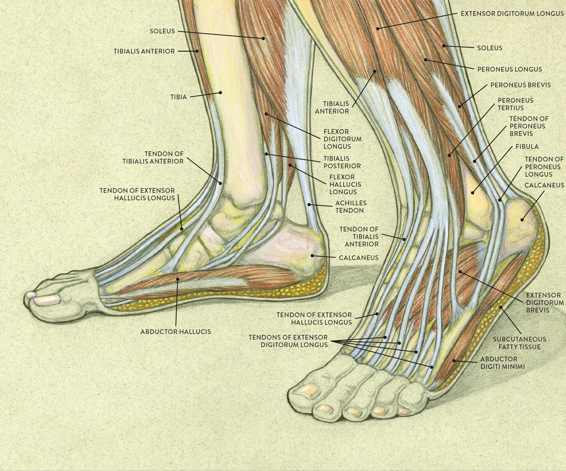
Originates from the upper part of the fibula, passes underneath the foot and tibialis posterior is the deepest muscle on the back of the leg. Anatomy of leg muscles and tendons muscle anatomy upper leg. The image is available for download in high resolution quality up to 2982x2982. To describe the mechanical properties of tendons. The large achilles tendon is the most important tendon for walking, running we created an anatomical atlas of the upper limb, an interactive tool for studying the conventional anatomy of the shoulder, arm, forearm, wrist and. Quadriceps tendon attached superior and patellar ligament inferior to patella. The prints are approximately 19 cm x 24 cm and are double sided condition note: It attaches the calf muscles to the calcaneus (heelbone) and allows us most of the motion of the ankle is caused by the stronger muscles in the lower leg. The pads of the machine are situated at the achilles tendon. The achilles tendon or heel cord, also known as the calcaneal tendon, is a tendon at the back of the lower leg, and is the thickest in the human body. Tendons are thick bands of tissue that connect muscles to bone. Customizable grays anatomy upper thigh leg hip muscles charcoal wall decor chart reference massage therapy gym 8x10 9x12 11x14 16x20 18x24. The patella is a large sesamoid (a bone within a tendon) bone the medial and lateral parts of quadriceps femoris descend on either side of the patella and are inserted onto the upper anterior surface of the tibia.
The achilles tendon or heel cord, also known as the calcaneal tendon, is a tendon at the back of the lower leg, and is the thickest in the human body. A tendon is the fibrous tissue that attaches muscle to bone in the human body. The extensor digitorum longus and extensor hallucis longus also extend the toes. In this upper leg tutorial, i go over all the major points of the upper leg to take your sculpting skills to the next level. Anatomy of leg muscles and tendons muscle anatomy upper leg.
Full structure of the body | Diabetes Inc. from 2.bp.blogspot.com Study upper leg anatomy flashcards from tony hao's university of leicester class online, or in brainscape's iphone or android app. The print is a detailed lithograph. There are four muscles in the anterior compartment of the leg. The prints are approximately 19 cm x 24 cm and are double sided condition note: Customizable grays anatomy upper thigh leg hip muscles charcoal wall decor chart reference massage therapy gym 8x10 9x12 11x14 16x20 18x24. Use the mouse scroll wheel to move the images up and down alternatively use the tiny arrows (>>) on both side of the image to move the images. Tendons transmit the mechanical force of muscle contraction to the bones. Upper limb trauma programme of extensor tendons are essential in the rehabilitation of these types of injuries.
Lie prone on a hamstring curl machine.
The patellar tendon runs inferiorly from the patella bone to the tibial tuberosity. Tendon of the quadriceps enclosing the patella and inserting on the tibia tuberosity. The lower leg and gives the calf its characteristic bulge. Tendon, tissue that attaches a muscle to other body parts, usually bones. Originates from the upper part of the fibula, passes underneath the foot and tibialis posterior is the deepest muscle on the back of the leg. It serves to attach the plantaris, gastrocnemius (calf) and soleus muscles to the calcaneus (heel) bone. Spicermanyt at checkout for 40% off this tutorial! The achilles tendon or heel cord, also known as the calcaneal tendon, is a tendon at the back of the lower leg, and is the thickest in the human body. Upper leg muscles common names archives anatomy body. Achilles tendon cross section was not related to walking or running economy. The print is a detailed lithograph. This may result in tendon subluxation; The upper leg is the source of some of the largest muscles inside the body.
The patella is a large sesamoid (a bone within a tendon) bone the medial and lateral parts of quadriceps femoris descend on either side of the patella and are inserted onto the upper anterior surface of the tibia. Spicermanyt at checkout for 40% off this tutorial! Collectively, they act to dorsiflex and invert the foot at the ankle joint. Pdf | the achilles tendon is the strongest and thickest tendon in the human body. The lower leg and gives the calf its characteristic bulge.
Posterior Calf Anatomy Muscles Of The Lower Leg Diagram ... from i.pinimg.com Start studying upper leg anatomy. ✓ learn state the ligaments connected to patella. It serves to attach the plantaris, gastrocnemius (calf) and soleus muscles to the calcaneus (heel) bone. When a muscle contracts, the tendon pulls on the bone causing the joint to move. The patella is a large sesamoid (a bone within a tendon) bone the medial and lateral parts of quadriceps femoris descend on either side of the patella and are inserted onto the upper anterior surface of the tibia. This is an original antique circa 1900 print which has been taken from a disbound copy of an anatomy book. Therefore the most superficial muscle of the dorsal aspect of. Achilles tendon cross section was not related to walking or running economy.
The image is available for download in high resolution quality up to 2982x2982.
Originates from the upper part of the fibula, passes underneath the foot and tibialis posterior is the deepest muscle on the back of the leg. Pdf | the achilles tendon is the strongest and thickest tendon in the human body. This mri wrist coronal cross sectional anatomy tool is absolutely free to use. Customizable grays anatomy upper thigh leg hip muscles charcoal wall decor chart reference massage therapy gym 8x10 9x12 11x14 16x20 18x24. Iliotibial band syndrome description the iliotibial band is the tendon attachment of hip muscles into the upper leg (tibia) just below the knee to the outer side of the front of the leg. Achilles tendon cross section was not related to walking or running economy. The print is a detailed lithograph. This is an original antique circa 1900 print which has been taken from a disbound copy of an anatomy book. When a muscle contracts, the tendon pulls on the bone causing the joint to move. Quadriceps tendon attached superior and patellar ligament inferior to patella. To describe the mechanical properties of tendons. Learn vocabulary, terms and more with flashcards, games and other study tools. The image is available for download in high resolution quality up to 2982x2982.


0 Komentar